This lab investigates the effects of trauma on osteoarthritis at the cellular level. We are currently studying cell death, specifically necrosis and apoptosis, and researching possible membrane repair mechanisms. Our lab has also developed a bovine cartilage explant model. Here is a partial listing of the equipment available in this lab.
1. Laminar Flow Hood
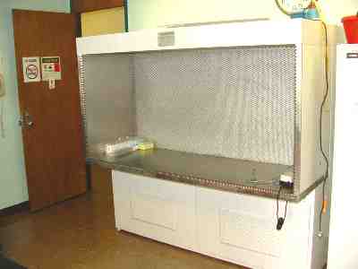
- Biosafety Level 1 Containment Protocol
- Provides optimal aseptic working environments
- Used for mammalian cell culture: bovine chondrocytes and cartilage tissue plugs
- Used for microbial cultures
- HEPA filters that have 99.9% efficiency on particles of 0.3um or more
- Seventy-two inch model
2. Water-Jacketed CO2 Incubator
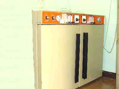
- Maintained at 37° C, 5% CO2, 95% humidity
- Temperature Range: 5-60 degrees Celsius (+0.2 degrees)
- Water Jacket keeps cultures warm during power failures
- Manually filled water pan has evaporative humidity up to 98% relative humidity
- Provides optimal environment for bovine cartilage tissue plugs
3. Microscopes & Digital Camera
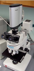
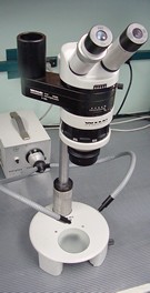
- Wild Heerbrugg Stereo Dissection microscope
- Leitz Dialux 20 fluorescent microscope
- Polaroid DMC2 digital camera optional attachment to either scope for high resolution microscopic photos
- Fluorescent light source and different microscope filters for various staining techniques
4. Rotary Microtome
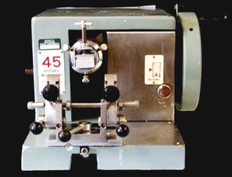
- Capable of slicing tissue specimens into thin slices for analysis
- Used in research for histology and to examine tibio-femoral explants
5. In Vitro Explant Loading Device
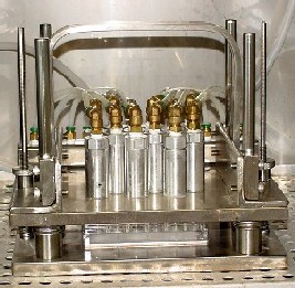
- Custom-designed pneumatic "cartilage exerciser"
- Biocompatible—operates within CO2 incubator—explants kept in standard tissue culture media in sterile well plates
- Adjustable & accurate—output loads and waveforms are controlled through a computer interface
- Simulates the mechanical loading environment of a joint