Jump to HeaderJump to Main ContentJump to Footer
Michigan State University
Center for Neuromusculoskeletal Clinical Research

Center for Neuromusculoskeletal Clinical Research

  • Home
  • |
  • About
  • |
  • Personnel
    • Klemont Caron
    • Jacek Cholewicki, PhD
    • Lisa DeStefano, DO
    • Jessica Epstein
    • Richard Hallgren, PhD
    • Angela Lee MPH
    • JAimee Lippert
    • Clarence (NIC) Nicodemus, DO, PhD
    • Christopher Pohlod
    • John M. Popovich, Jr., PhD, DPT, ATC
    • Jacob Rowan, DO
    • Mathew A Zatkin DO
  • |
  • Opportunities
  • |
  • Department of OMM
  • |
  • Contact
Center for Neuromusculoskeletal Clinical Research
  • Home
  • About
  • Personnel
  • Opportunities
  • Department of OMM
  • Contact

< Home

< About

< Personnel

  • Klemont Caron
  • Jacek Cholewicki, PhD
  • Lisa DeStefano, DO
  • Jessica Epstein
  • Richard Hallgren, PhD
  • Angela Lee MPH
  • JAimee Lippert
  • Clarence (NIC) Nicodemus, DO, PhD
  • Christopher Pohlod
  • John M. Popovich, Jr., PhD, DPT, ATC
  • Jacob Rowan, DO
  • Mathew A Zatkin DO

< Opportunities

< Department of OMM

< Contact

Center for Neuromusculoskeletal Clinical Research > Current Research >

Pediatric Skull Fracture Research

Currently, there is no reliable biomechanical model for pediatric skull fractures. The OBL has begun researching the material and mechanical properties of the developing porcine skull during impacts in order to better understand the nature of injury on developing cranial bone in humans.

The project, titled "A Forensic Pathology Tool to Predict Pediatric Skull Fracture Patterns," involves the OBL, MSU Forensic Anthropologist Dr. Todd Fenton. The collaborative effort from multiple departments hopes to generate a computational model for pediatric head injury that will be used by Forensic Pathologists in cases involving abuse.
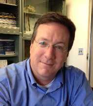
Dr Fenton

Dr. Todd Fenton, Ph.D.
Michigan State University

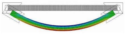
The OBL's preliminary studies investigated the material properties of the developing porcine skull. Through experimentation on thin beam specimens, our researchers hope to examine such properties as failure strength, energy absorbed and elastic modulus of developing cranial bone.
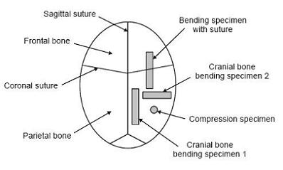
skull beams

beam bend

Top: Diagram of beam specimens used to calculate material properties.

Bottom: Computational model of beam specimen during bending.

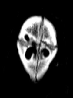
The research at the OBL will also include impact testing on cranial specimens. CT Scans will be done at Wayne State University after impact testing in order to gain a detailed view of fractures on the cranial surface. The mechanics of the skull fracture propagation will then be examined in order to generate the final computational model.
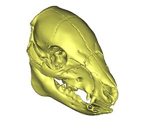
iso higher

Computer generated model of porcine skull.
ct scan

CT Scan of porcine specimen after impact.

Links

  • Osteopathic Manipulative Medicine

Resources

  • About
  • Opportunities

College of Osteopathic Medicine

  • About the College
  • Future Students
  • Our Mission
  • News

Contact us

517-353-9110

Center for Neuromusculoskeletal Clinical Research

A413 East Fee Hall 965 Wilson Road East Lansing, MI 48824

Follow Us

  • Visit our Facebook page
  • Visit our Instagram page
  • Visit our YouTube page

If you're having accessibility issues, please let us know.

  • Contact Information|
  • Site Map|
  • Privacy Statement|
  • Site Accessibility|
  • Call MSU: (517) 355-1855|
  • Visit: msu.edu|
  • Notice of Nondiscrimination|

SPARTANS WILL|© Michigan State University|