MSU's OBL has been performing research to develop efficient surgical methods and cutting-edge medical products. Below is a description of a couple recent studies conducted in the OBL.
Orthobiologic Material Development and Evaluation
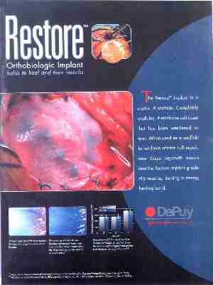
 The OBL recently assisted in the development of surgical procedure and material to repair a torn rotator cuff tendon (RCT). The procedure involved the utilization of a swine small intestine submucosa (SIS), developed at Purdue University. Our investigators have also co-authored the year 2000 O'Donoghue Award-winning paper that was recently published in the American Journal of Sports Medicine, titled "Tissue-Engineered Rotator Cuff Tendon Using Porcine Small Intestine Submucosa" (29(2):175-184) (Abstract). This manuscript described the basic science research conducted at MSU and the OBL that resulted in development of the Restore Orthobiologic Inplant DePuy Inc. This product was recently advertised in the American Journal of Sports Medicine and highlighted some of the biomechanical test data generated at the OBL.

Advertisement from the American Journal of Sports Medicine which shows biomechanical test data (see the lower right image) generated in the OBL study.
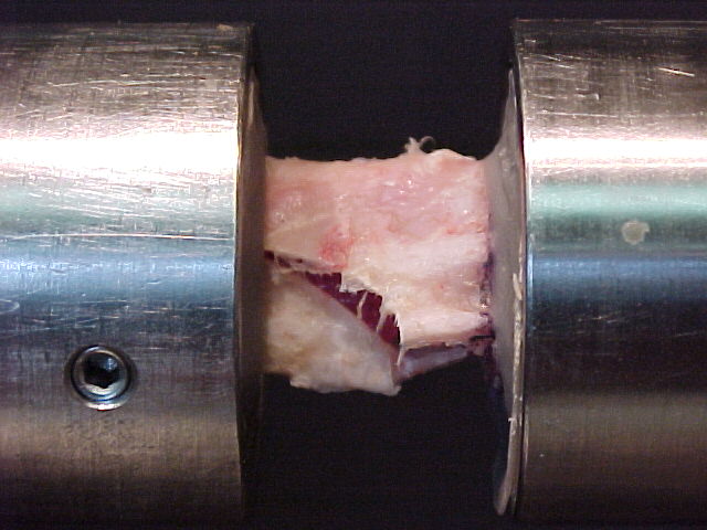
The OBL is currently evaluating several bone repair materials in studies with DePuy Inc. The studies, using the lapine tibial defect model, are shown at the right. Investigators have quantified the torsional strength of repaired defect sites over time and compared these strengths to those seen in normal and unrepaired tibias. Similar studies have been conducted at the OBL using the lapine radius, segmental defect model.

Torsional testing of the lapine proximal tibia.
Surgical Methods Evaluation
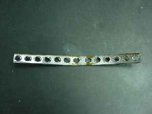
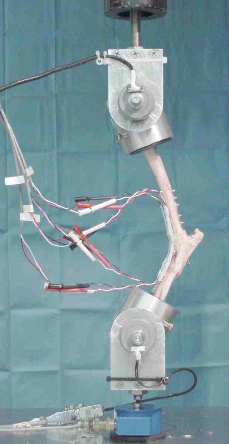
OBL investigators are also conducting experiments to develop more optimum surgical methods to improve the strength of surgical repairs. In a recent study, the OBL has worked with Dr. Loic DeJardin and the College of Veterinary Medicine to validate a surgical procedure developed at MSU to increase the fatigue strength of surgically repaired canine tibiotarsal joints. In this biomechanical study on the canine hindlimb preparation, the OBL used strain gauges mounted to the bone plates to test the efficacy of an added IM rod to this surgical procedure. Compressive loading of the construct verified an increase in fatigue life of the surgical procedure, via a reduction in plate strains and an increase in the structural stability of the hindlimb preparation with the addition of an IM rod into the tibia. These data are currently being prepared for publication in the veterinary orthopaedics literature.


Left: Tibiotarsal bone plate with a 130-degree bend. Right: The hindlimb construct mounted in the servo-hydraulic test machine with rotary encoders at the ends of the construct and a network of strain gauges on the plate.